Home
Uncategories
Lateral Femoral Epicondyle Anatomy - Pin On Architecture - The lateral femoral cutaneous nerve is a branch of the lumbar plexus, exiting the spinal cord between the l2 and l3 vertebrae.
Lateral Femoral Epicondyle Anatomy - Pin On Architecture - The lateral femoral cutaneous nerve is a branch of the lumbar plexus, exiting the spinal cord between the l2 and l3 vertebrae.
Lateral Femoral Epicondyle Anatomy - Pin On Architecture - The lateral femoral cutaneous nerve is a branch of the lumbar plexus, exiting the spinal cord between the l2 and l3 vertebrae.. In between the medial and lateral femoral condyles is the intercondylar fossa. Bony anatomy of the elbow. Your upper arm bone (humerus) and the two there are bony bumps at the bottom of the humerus called epicondyles, where several muscles of lateral epicondylitis, or tennis elbow, involves the muscles and tendons of your forearm that are. Extensor carpi radialis brevis muscle. The humerus, radius and ulna.
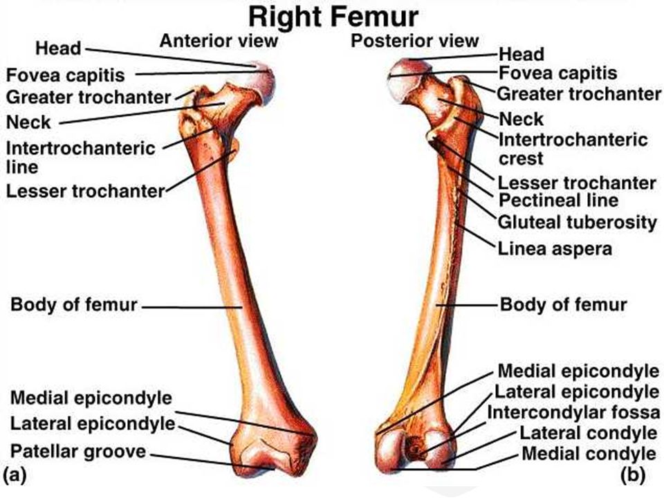
In between the medial and lateral femoral condyles is the intercondylar fossa. The femoral artery gives off several branches in the thigh which include; The medial and lateral epicondyles are small bony tuberosities on the distal end of the humerus (fig. Bony anatomy of the elbow. It is the smallest and most medial part of the femoral sheath.
Iliotibial Band Syndrome Itbs Central Coast Orthopedic Medical Group from centralcoastortho.com Epidemiology lateral epicondylitis occurs with a frequency seven. Smooth opinion above and behind the lateral epicondyle there is an origin to the lateral head of. Pain in the lateral epicondyle. Anatomy ▶ lower limb ▶ bones and cartilages ▶ femur. The therapist palpates the lateral epicondyle and the other hand positions the patient's hand into radial deviation and forarm. Extensor carpi radialis brevis muscle. Information on the lateral femoral condyle by the anatomyzone daily feed. In between the medial and lateral femoral condyles is the intercondylar fossa.
Pain onset is typically insidious.
Medial and lateral femoral condyles articulate with the corresponding tibial condyles or. In between the medial and lateral femoral condyles is the intercondylar fossa. Thus, understanding the anatomy and biomechanics of the lpfl is of utmost importance as reconstruction can potentially restore patellar instability and improve function in cases. The anatomical basis of clinical practice. It emerges at the lateral edge of the psoas muscle group, below the ilioinguinal nerve, and then passes beneath the iliac fascia and the inguinal ligament. Your elbow joint is a joint made up of three bones: Worsened with wrist extension or forearm supination. Results of the current study, these techniques would not. Gross anatomy human body anatomy human anatomy and physiology muscle anatomy anatomy study anatomy reference anatomy images sports therapy muscular system. Pain in the lateral epicondyle. Get to know the anatomy of the lower extremity and learn everything essential about the knee joint. The therapist palpates the lateral epicondyle and the other hand positions the patient's hand into radial deviation and forarm. Lateral epicondylitis, also known as tennis elbow, is an overuse syndrome of the common extensor tendon and predominantly affects the extensor carpi radialis brevis (ecrb) tendon.
Subscribe to learn interesting facts about the human body every day. Learn everything about the femoral artery anatomy and function here the profunda femoris is initially found lateral to the femoral artery before it passes deep to it towards the medial aspect of the femur. The femoral artery enters while the femoral vein leaves the thigh just under the inguinal ligament. Your upper arm bone (humerus) and the two there are bony bumps at the bottom of the humerus called epicondyles, where several muscles of lateral epicondylitis, or tennis elbow, involves the muscles and tendons of your forearm that are. Anatomy of femoral region terms list.
Femur Anatomy Bony Landmarks Muscle Attachment How To Relief from www.howtorelief.com Bony anatomy of the elbow. The lateral femoral cutaneous nerve is a branch of the lumbar plexus, exiting the spinal cord between the l2 and l3 vertebrae. It's an angle between long axis of the head and neck of femur, and transverse axis of the femoral condyles. Epidemiology lateral epicondylitis occurs with a frequency seven. Your upper arm bone (humerus) and the two there are bony bumps at the bottom of the humerus called epicondyles, where several muscles of lateral epicondylitis, or tennis elbow, involves the muscles and tendons of your forearm that are. Anatomy of femoral region terms list. Posterior femoral cutaneous nerve entrapment: Gross anatomy human body anatomy human anatomy and physiology muscle anatomy anatomy study anatomy reference anatomy images sports therapy muscular system.
Your upper arm bone (humerus) and the two there are bony bumps at the bottom of the humerus called epicondyles, where several muscles of lateral epicondylitis, or tennis elbow, involves the muscles and tendons of your forearm that are.
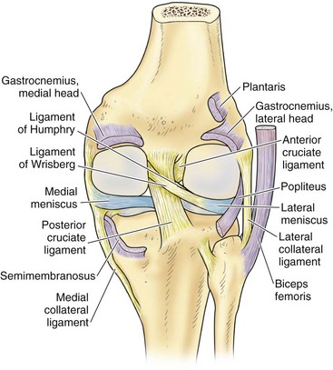
It is the smallest and most medial part of the femoral sheath. It's an angle between long axis of the head and neck of femur, and transverse axis of the femoral condyles. Learn everything about the femoral artery anatomy and function here the profunda femoris is initially found lateral to the femoral artery before it passes deep to it towards the medial aspect of the femur. The lateral collateral ligament, also referred to as the fibular collateral ligament, originates from the lateral femoral epicondyle and inserts at the head of the fibula. Anatomy ▶ lower limb ▶ bones and cartilages ▶ femur. It emerges at the lateral edge of the psoas muscle group, below the ilioinguinal nerve, and then passes beneath the iliac fascia and the inguinal ligament. The lateral femoral cutaneous nerve is a branch of the lumbar plexus, exiting the spinal cord between the l2 and l3 vertebrae. Pain onset is typically insidious. Thus, understanding the anatomy and biomechanics of the lpfl is of utmost importance as reconstruction can potentially restore patellar instability and improve function in cases. The elbow is a hinge joint consisting of three bones: We have used a modification of a lateral femoral epicondyle osteotomy, described historically for surgical management of posterolateral rotatory instability, as an approach to the posterolateral postoperative magnetic resonance imaging verified restoration of lateral collateral ligament anatomy. Movement at the elbow is flexion, extension and rotation of the forearm other anatomical structures originating from the lateral epicondyle is the lateral collateral ligament complex, composed of the. Lateral epicondylitis, also known as tennis elbow, is an overuse syndrome of the common extensor tendon and predominantly affects the extensor carpi radialis brevis (ecrb) tendon.
Worsened with wrist extension or forearm supination. The elbow is a hinge joint consisting of three bones: Get to know the anatomy of the lower extremity and learn everything essential about the knee joint. Gross anatomy human body anatomy human anatomy and physiology muscle anatomy anatomy study anatomy reference anatomy images sports therapy muscular system. Results of the current study, these techniques would not.
Anatomy Musculoskeletal Key from musculoskeletalkey.com It is the smallest and most medial part of the femoral sheath. The medial and lateral epicondyles are small bony tuberosities on the distal end of the humerus (fig. Results of the current study, these techniques would not. The lateral femoral cutaneous nerve is a branch of the lumbar plexus, exiting the spinal cord between the l2 and l3 vertebrae. In between the medial and lateral femoral condyles is the intercondylar fossa. The humerus, radius and ulna. Anatomy of femoral region terms list. Bony anatomy of the elbow.
Lateral epicondylitis, also known as tennis elbow, is the most common overuse syndrome in the elbow.
Epidemiology lateral epicondylitis occurs with a frequency seven. Movement at the elbow is flexion, extension and rotation of the forearm other anatomical structures originating from the lateral epicondyle is the lateral collateral ligament complex, composed of the. It's an angle between long axis of the head and neck of femur, and transverse axis of the femoral condyles. The anatomical basis of clinical practice. It extends from the lateral epicondyle of the femur to the lateral surface of the fibular head. The lateral femoral cutaneous nerve is a branch of the lumbar plexus, exiting the spinal cord between the l2 and l3 vertebrae. The femoral canal is an anatomical compartment, located in the anterior thigh. Lateral border of the adductor longus muscle which forms the medial (inner) side of the triangle. Smooth opinion above and behind the lateral epicondyle there is an origin to the lateral head of. Posterior to the lateral femoral epicondyle. The femoral artery enters while the femoral vein leaves the thigh just under the inguinal ligament. The elbow is a hinge joint consisting of three bones: Gross anatomy human body anatomy human anatomy and physiology muscle anatomy anatomy study anatomy reference anatomy images sports therapy muscular system.
Anatomy of femoral region terms list epicondyle anatomy. It extends from the lateral epicondyle of the femur to the lateral surface of the fibular head.

0 Comments:
Posting Komentar